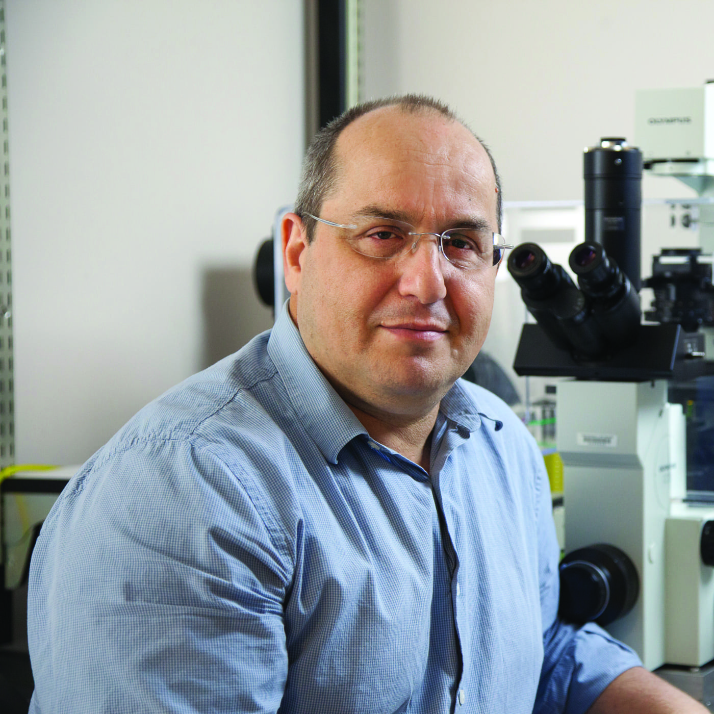
(April, 2024) Miami Project’s Dr. Pantelis Tsoulfas, Associate Professor of Neurological Surgery and Cell Biology, continues a long line of collaborative work with Dr. Murray Blackmore, Professor of Biomedical Sciences at Marquette University. Equipped with a new federal award, this accomplished pair continues to employ their established and proven neuroanatomical approach while venturing into new territory mapping the entirety of the supraspinal, or brain, connections between the central nervous system (CNS) and functional recovery after spinal cord injury (SCI).
At first glance it might not be obvious why the brain changes after SCI, since trauma occurred to the spinal cord, and it is in the cord that the glia and fibrotic scar forms. Considering the old adage “use it or lose it”—which is formalized in neuroscience as activity-based plasticity—it becomes clear that the reduction in sensory input from the body, no longer transmitted to the brain, leads to a reduction in brain regions that would typically process that input. Known as cortical remapping, these changes also occur not only in brain regions that receive information but also in outgoing regions that no longer routinely activate due to the futility of their no longer transmitted message to the body. Neuroscientists have long since studied the connection between select brain regions after SCI. Drs. Tsoulfas and Blackmore have been diligently pursuing their goal of charting the entire network of brain connections, termed the “supraspinal connectome”, rather than focusing on just one or two, to establish a neuroscientific framework for understanding the variations in functional outcomes after SCI. Their new award integrates previously proven but separate approaches in a pivotal and thorough investigation, demonstrating the power of the model they have developed.
Drs. Tsoulfas and Blackmore’s supraspinal connectome journey began in 2018, when Dr. Blackmore was a postdoctoral fellow at The Miami Project. Blackmore, and Tsoulfas embarked on implementing a novel viral technique in SCI that had been developed for labeling neurons projecting their fibers to various targets throughout the central nervous system. The strength of this viral method lies in its capacity to label all intact brain connections with just a single application of the tracer in the spinal cord. The difficulty in leveraging this capability lies in the requirement to image an entire, undamaged brain rather than just a subregion as conventional technologies allow for. Thus, the realization the supraspinal connectome paradigm is made possible by a significant advancement in microscopy known as light-sheet microscopy. Traditional microscopy requires the sectioning, or slicing, of thin wafer-like sections of a sample so that light can penetrate and thus illuminate the tissue and the scientist’s chosen labels.
In light-sheet microscopy, the sample is not sectioned but instead cleared, using a chemical process making the tissue transparent. A sheet of light is then passed through the entire sample, in this case the whole brain, to illuminate and reconstruct in 3D the intact tissue without the need to section. Combining the viral tracer with light-sheet makes good sense given the ability to label the entire CNS and then image the neurons with their processes without disturbing anything in the environment.
Their latest project goes beyond simply capturing images of the CNS post-SCI; it also aims to understand how changes in the connectome relate to functional outcomes. By adopting a connectome approach, Tsoulfas, Blackmore, and their team aim to link changes in brain regions above the injury site with the restoration of motor function, aiming to understand the relationship between the connectome and behavior. The variability in recovery post-SCI, how it is that some smaller injuries lead to profound deficits while larger SCIs result in relatively minor impairments, remains an important and unanswered question.
Incorporating an element of causation into their groundbreaking study, the team plans to deactivate certain brain regions implicated through imaging as being involved recovery after SCI. If silencing leads to the loss of a previously regained function, it will provide strong evidence of that region’s causal involvement in the function being studied.
This federally funded sustaining collaboration, which adopts a systems-based approach to develop a neuroscience paradigm specific to SCI, exemplifies The Miami Project’s ethos and Tsoulfas’s career-long dedication to understanding and eventually curing SCI. “Science thrives on reciprocity and collaboration of different teams,” Tsoulfas emphasizes, “and the value of long-term professional relationships cannot be overstated.”
Upon completion of their project, Tsoulfas, Blackmore, and their team will have developed a comprehensive CNS labeling and imaging approach that integrates the supraspinal connectome with functional recovery after SCI, this paradigm will also shed light into the complex relationship between the supraspinal connectome and functional outcomes post-SCI. This approach will be incorporated into future analyses of interventions and treatments, attributing effects to specific nuclei regardless of the of intervention type, whether involves devices, pharmaceuticals, or biologic treatments.

